Ankle Anatomy
Ankle Anatomy
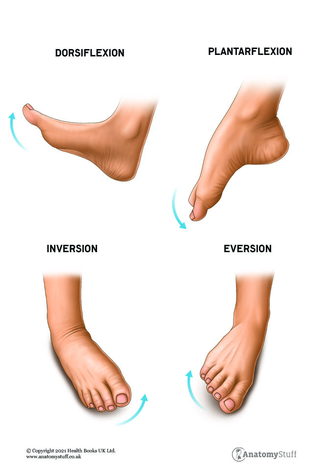
Three bones form the ankle joint: tibia, fibula and the talus. There are 4 main movements at the ankle: dorsiflexion, plantarflexion, inversion & eversion.
The joint is stabilized by three main groups of ligaments. The ligaments provide support on the medial (inside), lateral (outside) of the ankle and between the tibia and fibula bones. The ligaments prevent excessive motion, provide joint stability and act to create leverage for movement.
The anterior talofibular ligament provides support laterally. The ligament prevents the foot from inverting, or pointing inward. This ligament is typically injured when the ankle “rolls in”.
The interosseous membrane, anterior and posterior tibiofibular ligaments support the “upper ankle”. The membrane forms a web that spans the space between the tibia and fibular bones, whereas the tibiofibular ligaments are located closer to the ankle joint.
The posterior tibiotalar, tibiocalcaneal, tibionavicular and anterior tibiotalar ligament provide support medially. This group of ligaments collectively prevent the foot from everting, or pointing outwards. These ligaments are injured when the ankle “rolls out”. They are rarely injured since they are very strong.
There are four main groups of muscles that control the foot and ankle. The muscles located on the outside (lateral) and inside (medial) part of the lower leg control inversion and eversion, or the toes pointing inward or outward. The muscles on the front and back of the lower leg control dorsiflexion and plantarflexion.
The muscles work in synergy to provide effective and efficient movement.