Helping circus artists recover from low back pain: a movement system impairment style
If you are a circus artists (aerial, acrobat, contortionist or other performing artist) experiencing low back pain, this article is for you.
We will discuss why low back pain is so widespread in the circus artist community, how a physical therapists like me can help to identify a possible cause of your back pain, and what you can do to relieve yourself of low back pain.
Recreational circus has been a growing community in the past decade, with practitioners using performing arts as a form of exercise. And this is absolutely amazing! We need more folks participating in regular physical activity for physical and mental health- the circus arts are a perfect way to do it.
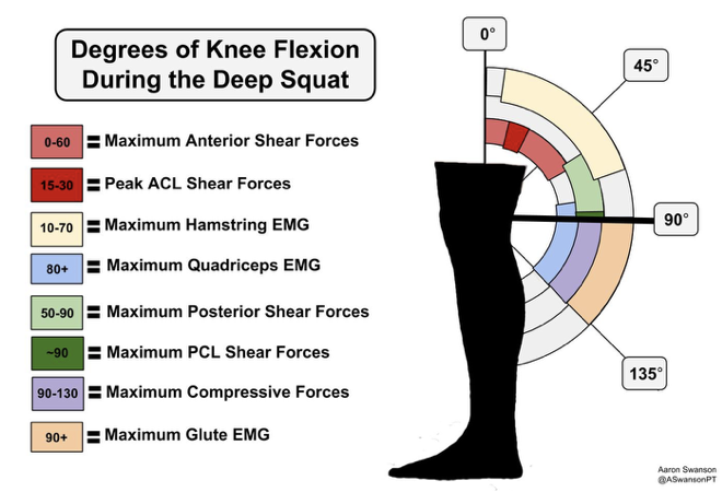
Low back pain is common in circus artists. Seeing that more people are participating in circus, this article may help prevent low back pain from occurring. One possible cause of low back pain in circus artists is the extremes of flexibility that you are required to have for “aesthetic” purposes. Great flexibility with strength can help you look in control of every inch of movement- appearing graceful. But repeatedly moving into these extremes of position may increase the risk of developing low back pain.
A 5 minute performance may require repeated spinal movements towards the end range of flexibility, oftentimes holding this position for several seconds. Over many years of practicing circus, you may find yourself favoring one side more than the other in certain positions or sequence of movements. The Movement System Impairment assessment we will now discuss is used to identify these imbalances in circus artists and direct treatment to correct them. This may help guide you in the right direction for assessing positions frequently performed in circus arts, with a trained eye.
Movement or positional imbalances often result in compensatory patterns or static position. These imbalances may help direct treatment towards the root cause: altered muscle function, areas of poor flexibility, pain or maybe even fear of the position itself.
The Movement System Impairment (MSI) assessment looks at a circus artists ability to maintain a specific body position with 8 different movements. Movement outside of, or inability to get into these positions will be a clinical observation that can then begin to establish a movement impairment diagnosis. The movements are chosen because they offer an opportunity to find a common movement-specific impairment, and begin there as a starting point for intervention. For example, if you have low back pain when inverting when hanging, and you also have low back pain when performing a leg lift with the MSI assessment, that would be the starting point for pain-provoking positions.
The MSI diagnosis contains 5 categories. The artist is categorized based on the direction preference of the limbs or spine in any of the 8 movements. Whens the movement assessment is over, the physical therapists will make a MSI-specific diagnosis, and a treatment plan will be developed with the artist.
Like every other single athlete out there, circus artists do not like to take time off. Every athlete knows that the more time spent on their craft gets them one step closer to their goal. It doesn’t matter if they’re fitness goals, technique acquisition, training for a performance, or just for peace of mind, athletes want and deserve to have a body that moves well.
If you are a circus artist experiencing low back pain and feel like you do not have a reliable source of direction, give our office a call and we will do whatever it takes to get you the help you need no matter where you are.
I also help circus artists improve their strength and flexibility with my circus strength training program. These programs are custom made, based on your specific goals and time. We first take a thorough history and examination, discussing previous injuries, how they’re being managed, and then coming up with a plan to get you swinging in the right direction.
Give our office a call and we can schedule a phone call to see if we’re a right fit for each other.